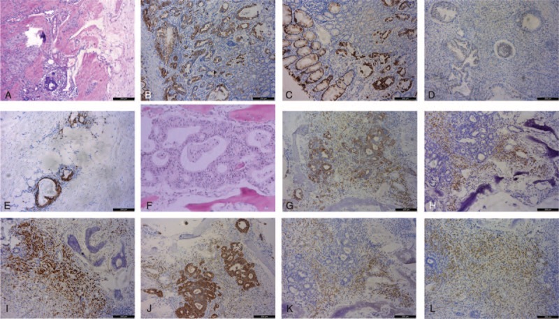
FIGURE 2.

Hematoxylin and eosin (H&E) staining (A and F, 100×) and immunohistochemical staining (B, C, D, E, G, H, I, J, K, and L, 100×) of the primary gastric carcinoma (A–E) and MP (F–L). Primary gastric cancer was ulcerative gastric adenocarcinoma with poor to moderate differentiation, histological grade II, and pT4aN0M0 stage. The tumor was immunopositive for CK7 (B), CK20 (C), CDX-2 (D), and villin (E). MGCP revealed poorly differentiated atypia adenocarcinoma cells (F), was immunopositive for CK7 (G), CK20 (H), CDX-2 (I), and villin (J), and was also immunopositive for CgA (K) and Syn (L). H&E = hematoxylin and eosin; MGCP = metastatic gastric cancer in the pituitary; MP = metastasis in the pituitary.
