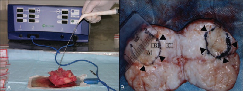
FIGURE 1.

MSI S-500 radiofrequency ablation apparatus and the procedures using which the fibroids were ablated. (A) MSI S-500 radiofrequency ablation apparatus was used to ablate the resected uterus. (B) The circle showed the scope of the ablated lesion, the diameter of the ablated lesion was 2.5 cm. “A” represents the center of the ablated lesion, “B” represents the edge of the ablated lesion, and “C” represented the area 1 cm away from the edge of the ablated lesion.
