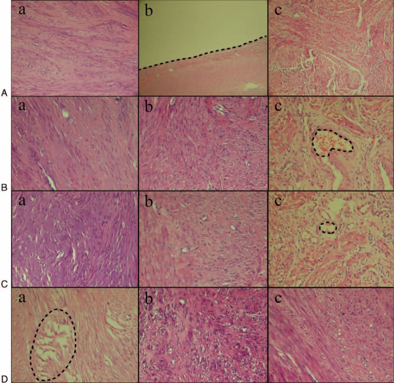
FIGURE 2.

Histological changes in the fibroid treated at different temperatures (×200). (A) Histological change of fibroid ablated in vitro at a temperature of 60°C. (a) Focal coagulation necrosis, with part of the muscle fibers being visible. (b) Bleeding line around the edge of the ablated lesion (the black line). (c) No degeneration, necrosis, or inflammatory cell infiltration in the fibroid tissue. (B) Histological changes in the fibroid ablated in vivo at a temperature of 80°C. (a) Coagulation necrosis, damaged muscle fiber structure, karyoclysis, and reduced nuclear color. (b) Lesser tissue necrosis compared with group A, damaged muscle fiber structure, but many cell nuclei visible. (c) No muscle cell necrosis and inflammatory cells filtration. There was a patent blood vessel in the center of lesion, with presence of red blood cells (the black circle). (C) Histological change in fibroid when ablated in vitro at a temperature of 100°C. (a) Coagulative necrosis, damaged muscle fiber structure, and karyorrhexis. (b) Partial regional necrosis, which was lesser compared with the group A. (c) No obvious fibroid cells degeneration, necrosis, but several small patent blood vessels with red blood cells (the black circle). (D) Histological change in fibroid when ablated in vivo at a temperature of 100°C. (a) Coagulative necrosis, electric shock like fracture, damaged muscle cell structure, and karyorrhexis (the black circle). (b) Focal coagulative necrosis, the extent of which was lesser compared with group A. (c) No obvious degeneration, necrosis, and inflammatory cells infiltration in the leiomyoma cells.
