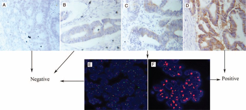
FIGURE 1.

Representative immunohistochemical (IHC) staining and fluorescence in situ hybridization (FISH) for HER-2 in rectal cancer cells (400×): (A) nonstaining is observed (0); (B) faint staining is observed in >10% of tumor cells (1+); (C) moderate staining is observed in >10% of the tumor cells (2+); (D) strong staining is observed in >10% of the tumor cells (3+); (E) tumors without HER-2 amplification; and (F) tumors exhibited HER-2 amplification with an HER-2/CEP17 ratio >2.0. FISH = fluorescence in situ hybridization, HER-2 = human epidermal growth factor receptor-2, IHC = immunohistochemical.
