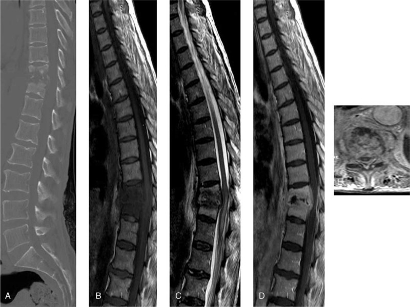
FIGURE 1.

Preoperative 3D CT scan (A) and sagittal T1- and T2-weighted MRI (B, C) showing a destruction T10–11 disc space and vertebral bodies. Gadolinium enhancement sagittal and axial T1-weighted images reveal epidural abscess and spinal cord compression.CT = computed tomography, MRI = magnetic resonance imaging.
