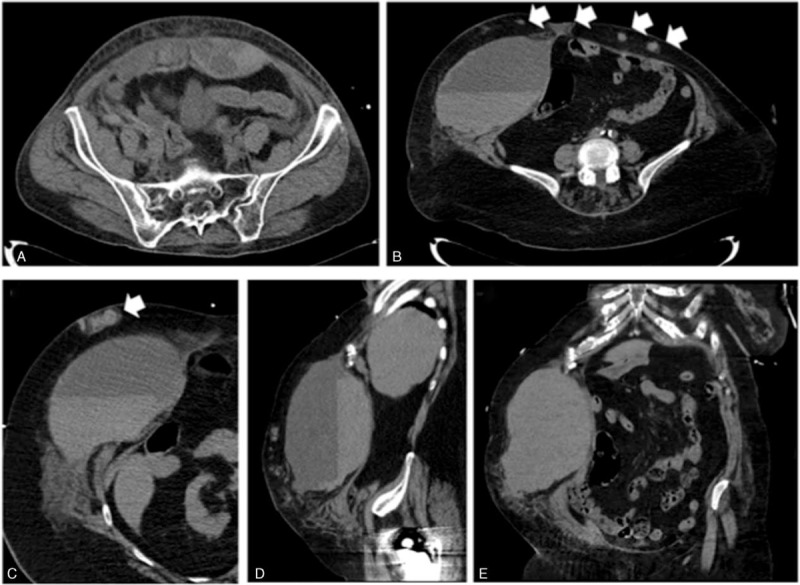
FIGURE 1.

Computed tomography (CT)-scan imaging of bleeding in the anterior abdominal wall. (A) Bleeding in the rectus abdominis muscle. (B, C) Horizontal sections showing an important bleeding in the right rectus abdominis muscle associated with several subcutaneous hematomas (white arrows), sagittal section (D), and coronal section (E).
