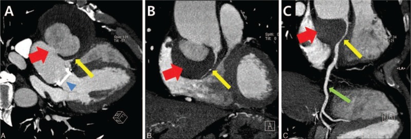
Figure 2.

Cardiac MDCT 7 years after aortic valve replacement. (A and B) 2-dimensional coronal cross-section images demonstrate a large saccular aneurysm of the ascending aorta just distal to the previously replaced prosthetic valve (79.7 × 72.8 mm in size). It appears to contain mural thrombi or hematoma and is seen here compressing the proximal RCA. (C) Curved MPR image demonstrates the compressed proximal RCA by the large aneuysmal sac with mural low-density opacities suggesting thrombi or hematoma. ∗ aortic aneurysm with mural thrombi or intramural hematoma (red arrows); eccentrically compressed proximal RCA (yellow arrows); normal mid-RCA (green arrow); prosthetic aortic valve (blue arrow head). MDCT = multidetector computed tomography, MPR = muliplanar reformatting, RCA = right coronary artery.
