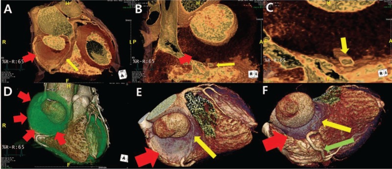
Figure 3.

Cardiac MDCT 7 years after aortic valve replacement, (A–D) 3-dimensional volume rendering images demonstrate the saccular type of aneurysm with a narrow neck. The proximal RCA is externally compressed by theaneurysmal sac with mural thrombi or hematoma on endoluminal or thrombotic setting. (E and F) Normal mid-to-distal RCA. MDCT = multidetector computed tomography, RCA = right coronary artery. ∗aortic aneurysm with mural thrombi or intramural hematoma (red arrows); eccentrically compressed proximal RCA (yellow arrows); normal mid-RCA (green arrow); prosthetic aortic valve (blue arrow head).
