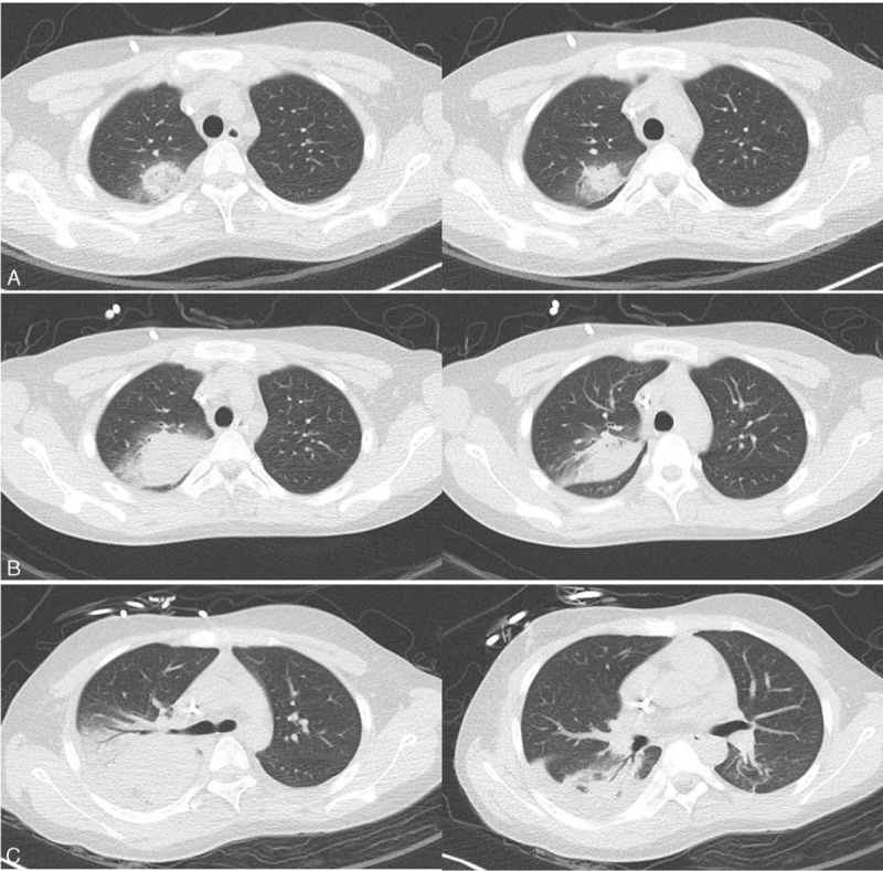
Figure 2.

Chest computed tomography showing pneumonic consolidation with surrounding ground glass opacity on the right upper lobe during the previous hospitalization (A), and aggravation of the pneumonic consolidation on fever days 6 (B) and 21 (C), respectively.
