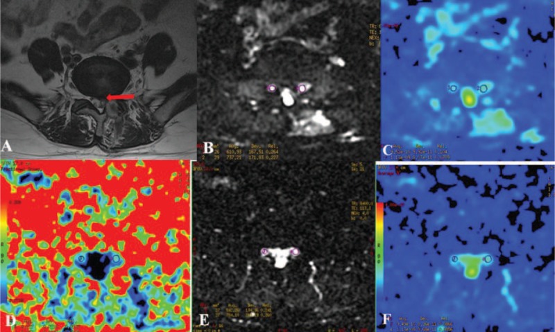
Figure 1.

The FA and ADC measurements before and after surgery in patients with unilateral S1 disc herniation. The S1 nerve root was compressed on the left side (A). Bilateral nerve roots were assessed by DTI before (B) and after surgery (D). The ADC (C and F) and FA (E) values at the most compressed part are measured using the manual setting of the ROIs before and after surgery. B, FA before surgery (left side: 0.187, right side: 0.262); E, FA after surgery (left side: 0.242, right side: 0.259); C, ADC (10−3 mm2/s) before surgery (left side: 1.451, right side: 1.311); F, ADC (10−3 mm2/s) after surgery (left side: 1.286, right side: 1.372). ADC = apparent diffusion coefficient, DTI = diffusion tensor imaging, FA = fractional anisotropy, S1 = sacral 1.
