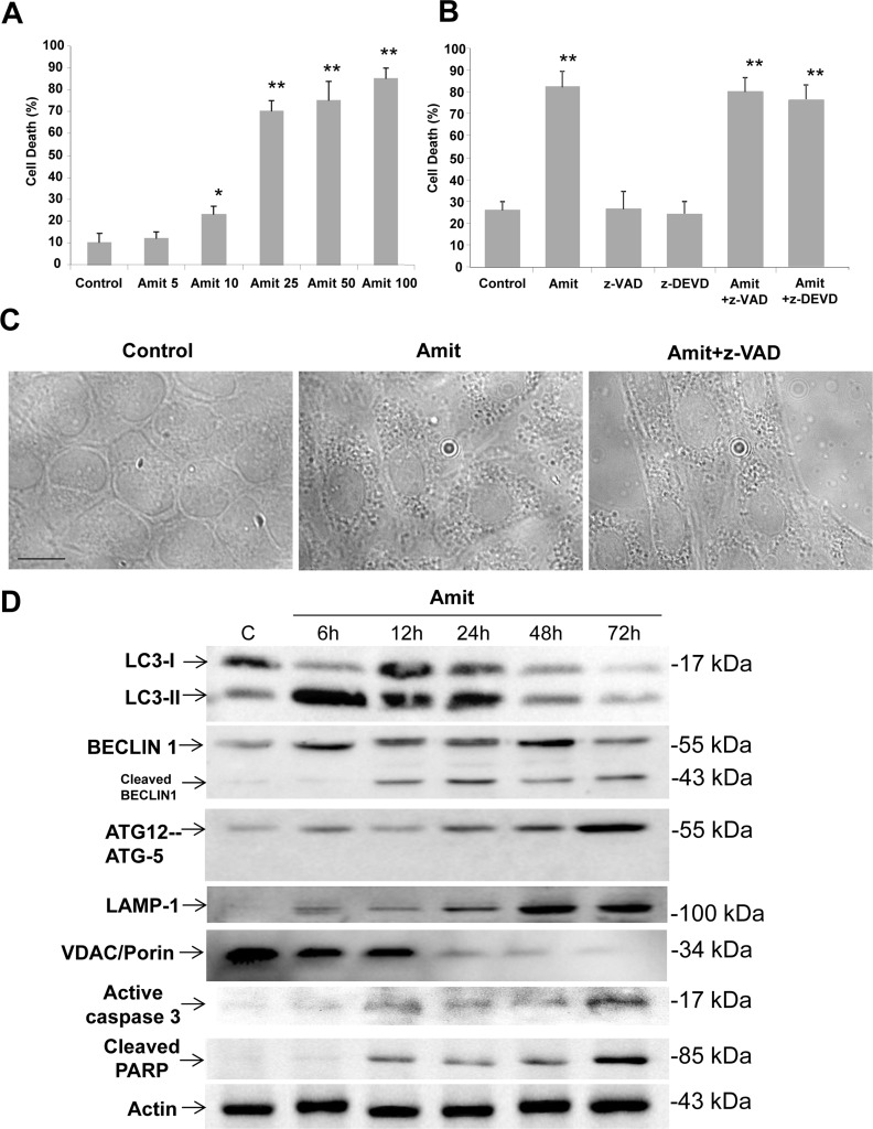
Figure 1. Amitriptyline reduces HepG2 cell viability.
A. Cells were seeded in six-multiwell plates, at a density of 100,000 cells/well. After 24 h of culture, serial concentrations of Amitriptyline (Amit) (0, 5, 10, 25, 50 and 100 μM) were added to the culture medium and cells were further incubated for 24h. Cells were then harvested and viability was analyzed by using the vital dye exclusion assay as described in the Materials and methods section. B. Caspase inhibition does not prevent Amitriptyline-induced cell death. HepG2 cells were treated with 50 μM Amitriptyline in the presence of z-VAD (50 μM) or z-DEVD (50 μM) for 24h. Cells were then harvested and viability was analyzed as described in the Materials and methods section. C. Phase-contrast light microscopy of HepG2 cells treated with Amitriptyline. Control cells, not exposed to Amitriptyline, showing no vacuolation. Cells exposed to 50 μM Amitriptyline for 6 hours, showing vacuolation. The treatment with 50 μM z-VAD did not prevent Amitriptyline induced vacuolation. D. Amitriptyline induces autophagy and apoptosis in HepG2 cells. Expression levels of protein markers of autophagy (LC3, BECLIN 1 and ATG12-ATG5), lysosomes (LAMP-1), mitochondria (VDAC/Porin) and apoptosis (active caspase 3 and cleaved PARP) were examined in HepG2 cells treated with 50 μM Amitriptyline for 6, 12, 24 and 48 hours by Western blotting. Actin was used as loading control.

