. 2016 Oct 11;5:e15461. doi: 10.7554/eLife.15461

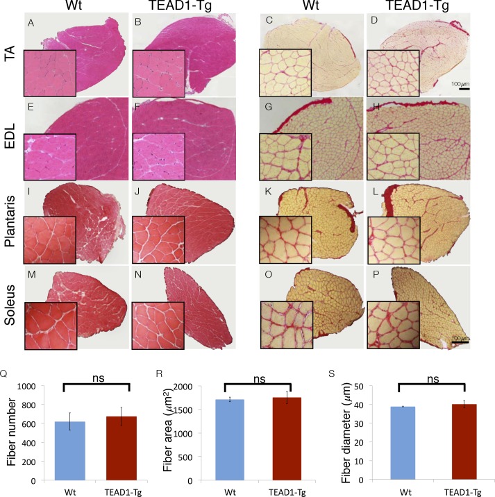
Figure 1. Skeletal muscle is histologically indistinguishable between Wt and TEAD1-Tg mice.

(A–P) H and E (AB, EF, IJ, MN) and Sirius Red (CD, GH, KL, OP) stains of TA (AD), EDL (EH), Plantaris (IL), and Soleus (MP) from Wt (A, C, E, G, I, K, M, O) and TEAD1-Tg mice (B, D, F, H, J, L, N, P). Insets are magnified images of the representative areas within the tissue. (QS) EDL fiber number (Q), fiber area (R), and fiber diameter (S) are quantified for both genotypes (n > 3 adult mice for all measurements).

DOI: http://dx.doi.org/10.7554/eLife.15461.003

Figure 1.

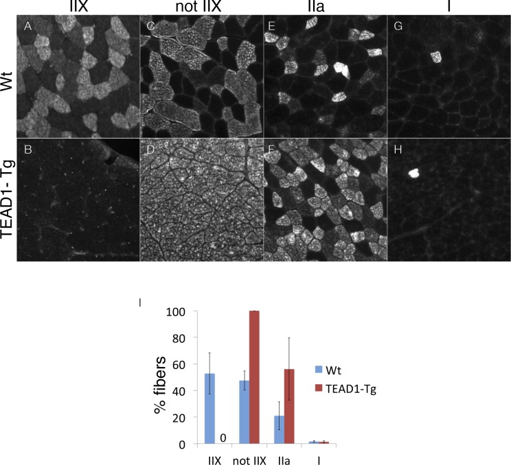
Figure 1—figure supplement 1. Ratios of type II fibers are changed in TEAD-Tg muscle.

Figure 1—figure supplement 1.

Wt (A, C, E, G) and TEAD1-Tg (B, D, F, H) TA muscles were queried for percentages of type IIX fibers (A, B), fibers that were not IIX (C, D), type IIa fibers (E, F), and type I fibers (G, H). The TA muscle was chosen for analysis because it is comprised of all muscle fiber types. The results were then quantified (I). n > 3 adult mice for all measurements, p<0.05 represented by (*), p<0.005 represented by (**), p<0.0005 represented by (***).