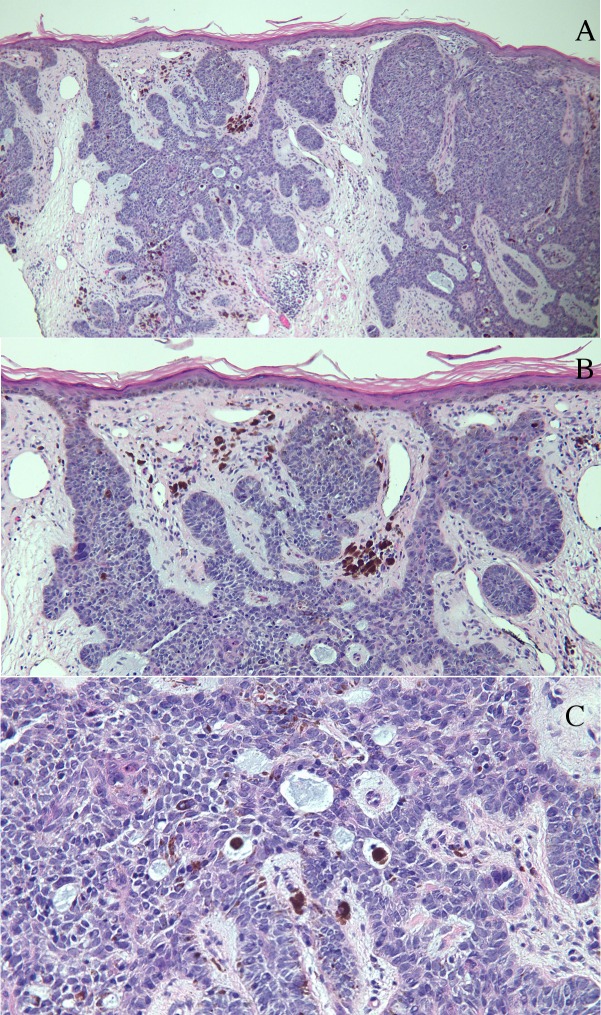
Figure 2. Umbilicus punch biopsy histology.
Low (A) and higher (B and C) magnification views of a 3 mm punch biopsy of the umbilical plaque in the woman from Figure 1. Microscopic examination showed nodular aggregates of basaloid tumor cells extending from the epidermis into the dermis (A). In the tumor and surrounding stroma, there were deposits of melanin, some of which were present in melanophages (B and C). Mucin, with or without melanin-containing melanophages, is present within the tumor aggregates (C). (hematoxylin and eosin stain: a = x4, b = x10, c = x40).

