Figure 5.
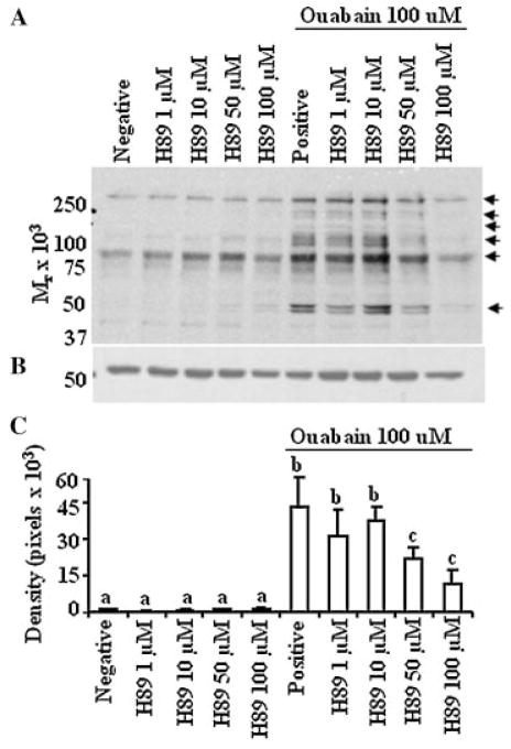
Tyrosine phosphorylation induced by ouabain was regulated by PKA in a dose-dependent manner. A: Bovine sperm preparations were processed and incubated as described in Figure 4, except that various concentrations of H89 (inhibitor for PKA) were used as the inhibitors. Arrows at the right of the immunoblot identify proteins exhibiting a change in the content of tyrosine phosphorylation; the results are representative of three experiments, using sperm preparations from three bulls. B: immunoblot was stripped and reprobed with anti β-tubulin to ensure equal loading between wells. C: Densitometry analysis on the effect of H89 on the content of phosphotyrosine in sperm treated with ouabain. A representative band (50 kDa) from blot A was quantified. In the absence of differences, densitometry data were directly compared between treatment groups. The results are representative of three experiments using sperm preparations from different bulls. Values presented are the mean ± SD. a–cValues without a common superscript differed (P <0.05).

