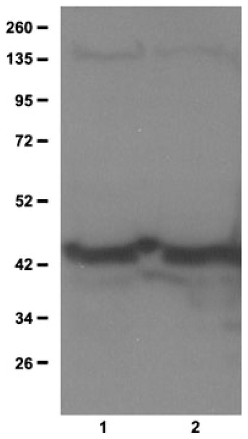
Figure 3.

Glutamate–ammonia ligase protein expression in testis and interstitial cells. Western blot analysis was done using anti-GLUL antibodies. Lane 1, total testis; lane 2, interstitial cell fraction. Molecular sizes of marker proteins are indicated on the left.
