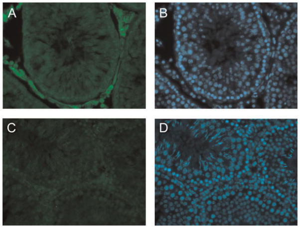
Figure 4.
Expression of glutamate–ammonia ligase in testis. Detection of GLUL protein expression in testis by immunofluorescence microscopy. Rat testes were frozen in OCT and sectioned. Testis sections were stained with an anti-GLUL antibody followed by a secondary antibody conjugated with Cy3. Panel A: Sections were stained using anti-GLUL antibody. Panel B: The same section was stained with DAPI. Panel C: Sections were stained using anti-GLUL antibody that was first blocked with GST–GLUL recombinant protein (negative control). Panel D: The same section was stained with DAPI.

