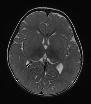
Fig. 2.

Magnetic resonance imaging (MRI) scan at 17 months of age revealed delayed but interval myelination associated with abnormal signal intensity of the bilateral thalami presenting as T2 hyperintensity of the posterior thalami in the region of the pulvinar nuclei (arrowhead) and T2 hypointensity in the anterior thalami
