
Fig. 4.
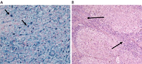
(a) Iron staining on liver resection specimen from case 2 (arrows: iron deposits in Kupffer cells). (b) Fibrosis on liver resection specimen from case 2 (arrows: fibrous septum running from the upper left portal tract to the lower right portal tract)