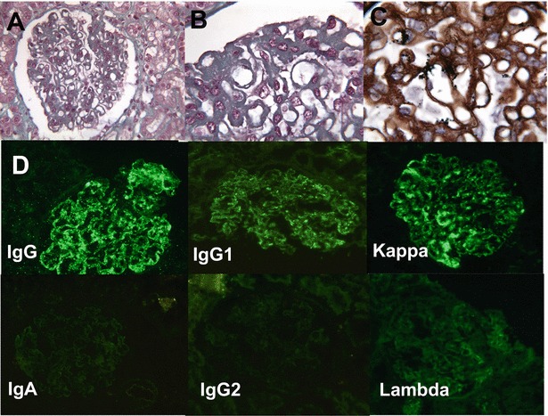
Fig. 2.

Patient 5, second kidney biopsy, membranoproliferative glomerulonephritis: (a) mesangial proliferation, extramembranous deposits (trichrome Masson, ×100). (b) Intramembranous and subendothelial deposits (trichrome Masson, ×250). (c) Irregular thickening of basal membranes, double contours (Marinozzi staining, ×250). (d) Immunofluorescence study (×100): Ig G, IgG1 and kappa light chain: intramembranous and extramembranous deposits. IgA, IgG2 and lambda light chain: no deposit; IgM, Ig3, Ig4 and fibrinogen: no deposit (data not shown)
