Figure 1.
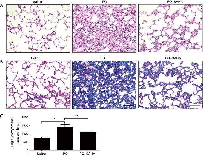
SAHA treatment lessens PQ-induced histological changes and hydroxyproline content in rat lung fibrosis. Histological sections of lung tissue were stained with H&E (A) and Masson trichrome (B). The images were captured under a light microscope. Original magnification, ×200. (A) Representative histological sections were stained by hematoxylin and eosin. Lung tissue sections of control animals showing normal lung morphologies: thin lined interalveolar septa with well-organized alveolar space; lung tissue sections of PQ-induced animals showed distorted lung morphologies: collapsed alveolar spaces with inflammatory exudates, wider and thickened interalveolar septa; lung tissue sections of SAHA treated PQ-induced animals: lower inflammatory infiltrates with lessened alveolar thickening; (B) effects of histopathological changes of PQ-induced lungs with Masson’s Trichrome stain: lung tissue sections of control animals with normal lung morphologies: scarcely deposited collagen in the lung parenchyma; lung tissue sections of PQ-induced animals showing dense collagen accumulations: collagen accumulations between alveoli; lung sections of SAHA treated PQ-induced animals showing reduced collagen depositions: reduced alveolar thickening with meager collagen; (C) effects of SAHA on the hydroxyproline content in the lungs of PQ-induced pulmonary fibrosis rats. Values are given as mean ± SD, n=8/group, ***P<0.001.

