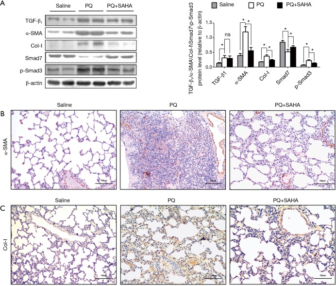
Figure 2.
SAHA supressed PQ-activated TGF-β1/Smad signaling. Rats were analyzed at 28 days after PQ intraperitoneal administration in the presence or absence of SAHA administered by gastric gavage. (A) Protein expression was analyzed by Western blotting with specific antibodies against TGF-β1, α-SMA, collagen I, Smad7 and Phospho-Smad3. Relative expression levels from samples were normalized by β-actin. Data are representative of three independent experiments with similar results. Values are given as mean ± SD, n=8/group, nsP >0.05, *P <0.05; (B) the distribution of lung α-SMA protein expression detected by immunohistochemistry in different groups (scale bar =100 µm). The images were captured under a light microscope (×200). Brown (α-SMA) stained lung sections images showing transdifferentiation of fibroblast into myofibroblast involved in the PQ-induced pulmonary fibrosis process. Lung segments of saline group animals showing localization predominantly around the interstitial space of the alveolar duct. In PQ-induced segments, α-SMA positive cells were observed in remodeled fibrotic alveoli. In SAHA treated segments, α-SMA positive cells decreased compare to PQ-induced segments; (C) the distribution of lung collagen I (Col-I) protein expression detected by immunohistochemistry in different groups (scale bar =100 µm). The images were captured under a light microscope (×200). Brown (collagen I) stained lung sections showing the degree of deposition of collagen fibers in lung tissue. Treatment with SAHA effectively prevented the accumulation of collagen I compare to PQ-induced segments.

