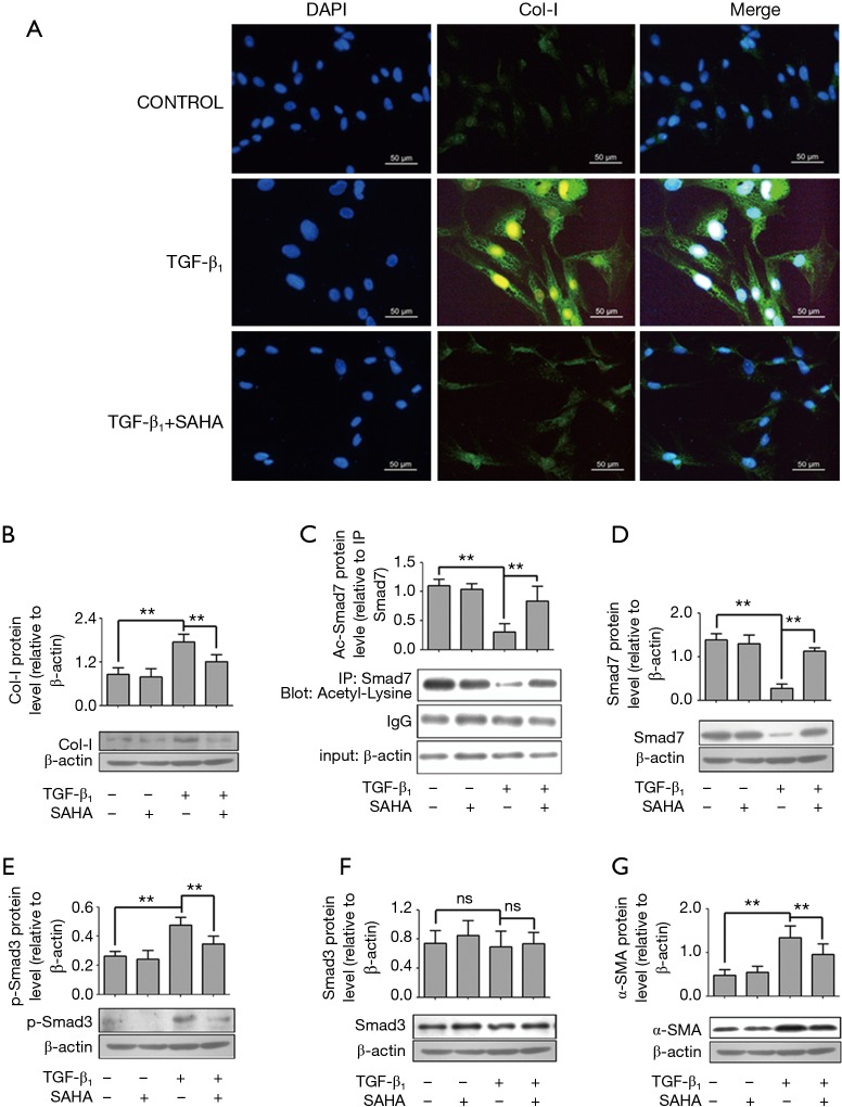
Figure 3.
SAHA inhibits TGF-β1-induced myofibroblast transdifferentiation via regulating Smad7 acetylation. (A) SAHA inhibited TGF-β1-indcued Collagen-I expression. HFL1 cells were incubated with 5 µM SAHA and 5 ng/mL TGF-β1 for 48 h. Cells were stained with anti-Col-I antibody and nucleus was stained with DAPI. Immunofluorescent studies demonstrated reduced intracellular collagen I staining (Figure 3A, green) after treating TGF-β1 cells for 48 h with SAHA. The images were visualized by immunofluorescence microscopy (×400); (C) the HFL1 cells were treated with or without SAHA (5 µM) and incubated with or without 5 ng/mL TGF-β1 for 48 h. Smad7 was immunoprecipitated from the samples. Acetylation of Smad7 was analyzed by immunoblotting using a rabbit anti-acetyl-lysine antibody; (B,D-G) The HFL1 cells were treated with or without SAHA (5 µM) and incubation with or without 5 ng/mL TGF-β1 for 48 h. Protein level was assessed by Western blotting with specific antibodies against Smad7 (D), Phospho-Smad3 (E), Smad3 (F), α-SMA (G) and collagen I (B). Expression was normalized to β-actin. Data are representative of three independent experiments with similar results. Values are given as mean ± SD, n=3/group, nsP >0.05, **P <0.01.

