Figure 1.
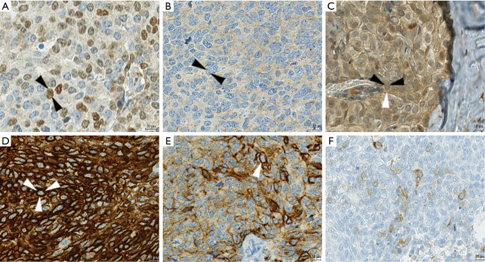
Examples of immunohistochemical staining. (A,B) In a case of basaloid TC, the primary tumor showed scattered nuclear expression of p40 (dark brown, black arrows mark the outline of one nucleus) whereas the relapse specimen of the same patient was negative for p40; (C) p21 was evaluated for nuclear and cytoplasmic staining separately; in this case of a B3, p21 was negative in the nucleus (black arrows), but moderately positive in the cytoplasm (white arrow); (D) Bcl-2, an oncogene often positive in TCs, shows strong cytoplasmic expression in this case of squamous TC (white arrows mark the cell membrane of a tumor cell); (E) only one case of TC expressed podoplanin (white arrow marks the cell membrane), a marker for lymphatic endothelium which can also be positive in thymic epithelial tumor cells; (F) neuroendocrine markers are also expressed in non-neuroendocrine TCs, like in this case with weak cytoplasmic expression of synaptophysin in a basaloid TC. TC, thymic carcinoma.

