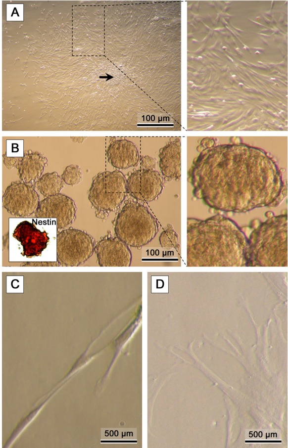
Fig. 3.

Representative micrographs of: (A) bone marrow MSCs. Formation of colonies (arrow) is typical in confluent MSC cultures (CFU-f). (B) Formation of Nestin-positive neurospheres (inner photo) after the first neurogenic induction step. (C, D) Disaggregated cells are directed into the second neurogenic induction stage yielding neurogenic phenotypes.
