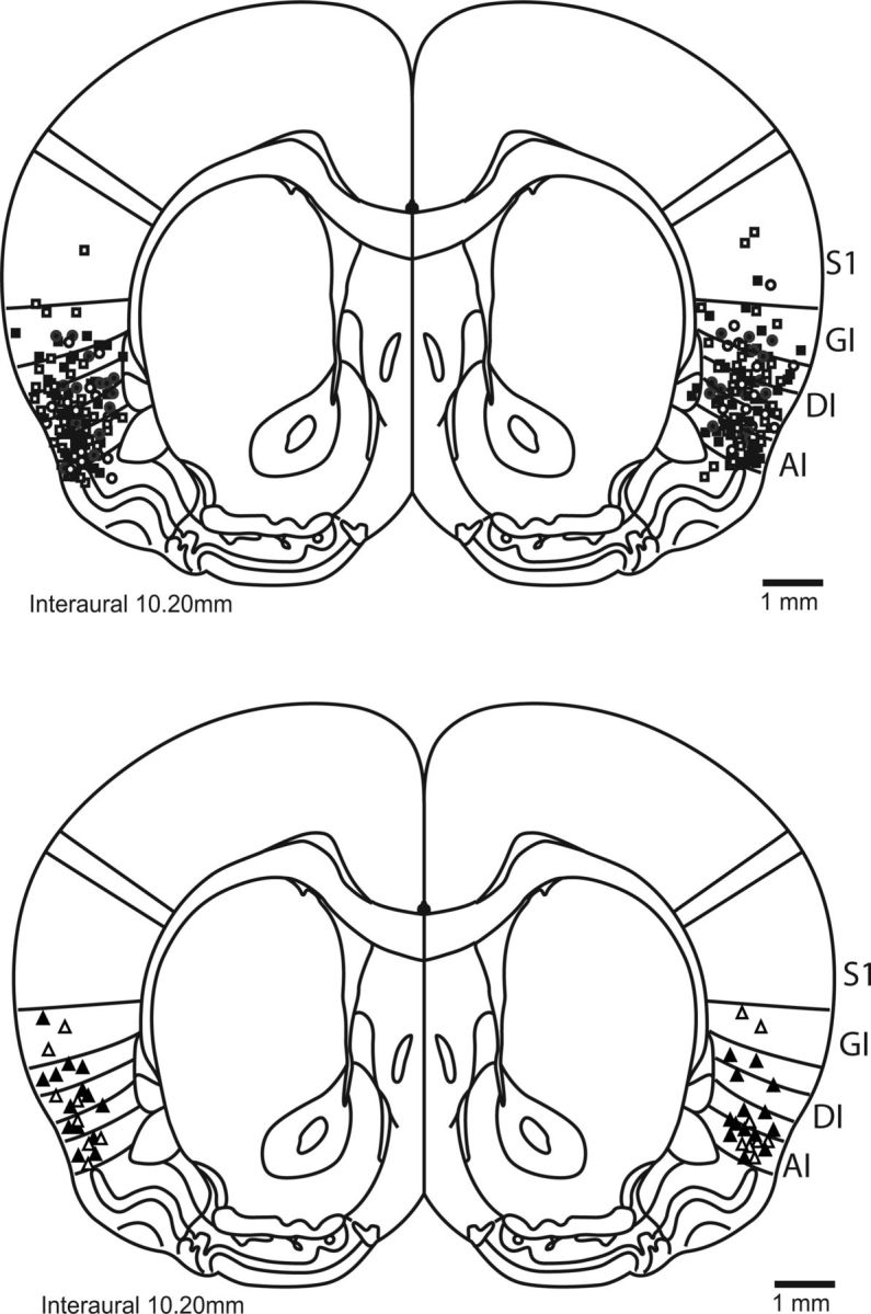
Figure 4.

Cannula placement in the rat GC. A schematic (reprinted with permission from Paxinos and Watson, 2007) of the rat brain at the level of GC (Katz et al., 2001) shows the locations of most infusions; rats in which infusions missed gustatory cortex (i.e., granular, agranular, and dysgranular insular) were excluded from analyses. S1, Somatosensory cortex; GI/AI/DI, granular/agranular/dysgranular insular cortex.
