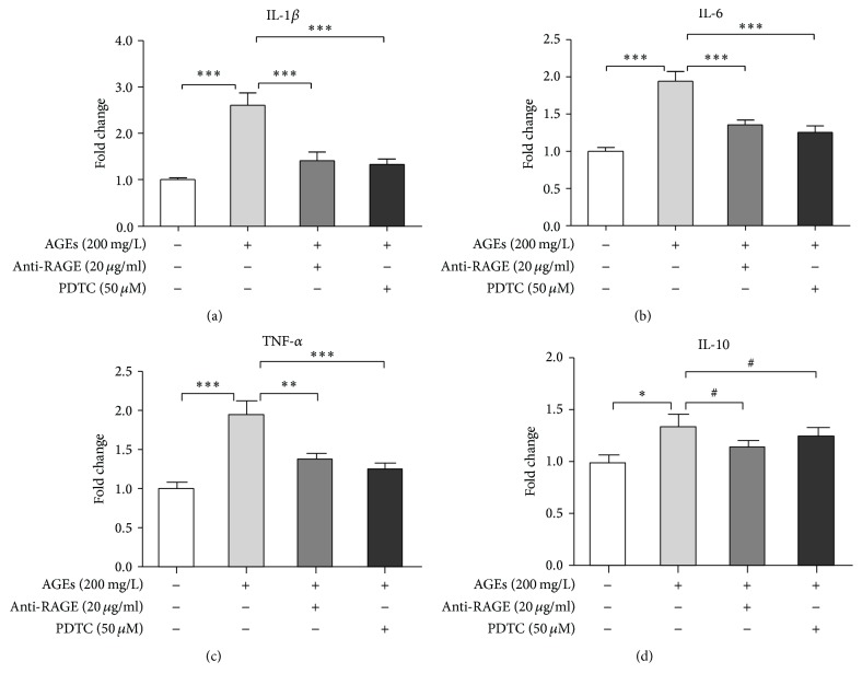
Figure 1.
AGEs-induced inflammatory response in BMDMs through RAGE/NFκB signaling. BMDMs were divided into 4 groups: control, AGEs, AGEs + anti-RAGE, and AGEs + PDTC group. Cells in the last two groups were pretreated with anti-RAGE antibody (20 μg/mL) or PDTC (50 μM) for 60 min, respectively, and then, together with AGEs group, the three groups were cultured with AGEs (200 mg/L) for 24 h. The control group was treated with BSA (200 mg/L) for the same amount of time. RNA then was extracted, and mRNA levels of IL-1β (a), IL-6 (b), TNF-α (c), and IL-10 (d) were measured by real-time PCR. Bar graphs represent the results (mean ± SD) of three independent experiments. One-way ANOVA was applied and all the overall ANOVA was significant. # p > 0.05; ∗ p < 0.05; ∗∗ p < 0.01; and ∗∗∗ p < 0.001 when compared between selected groups.

