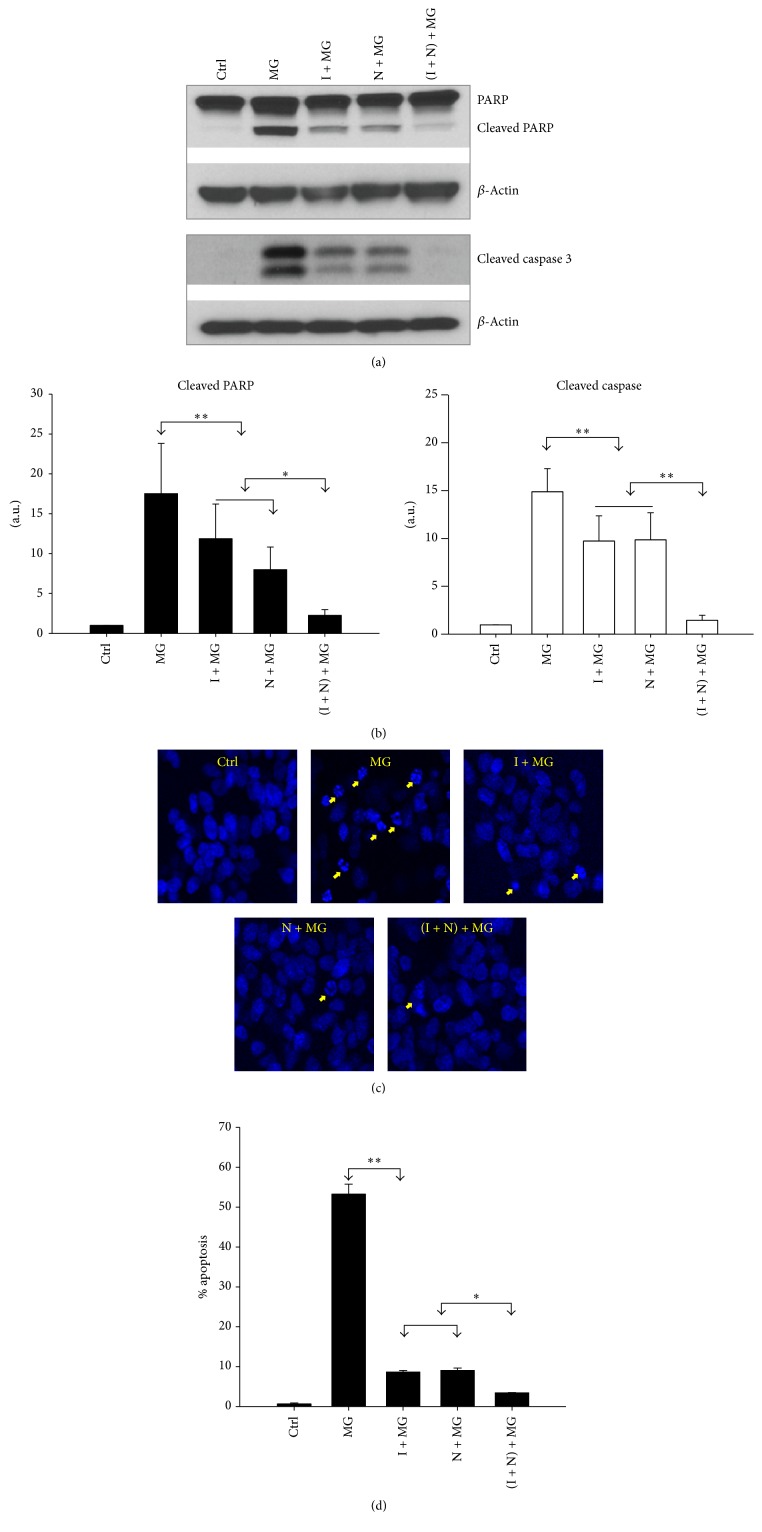
Figure 2.
IGF-1 and NAC repress MG132 induced apoptosis. (a) Western blot analysis for two apoptotic markers: cleaved PARP and cleaved caspase 3. Results are representative of three independent experiments. (b) Densitometric analysis from three separate experiments. β-Actin served as the loading control. (c) IGF-1 and NAC prevent MG132 induced nuclear morphological changes. Cells were pretreated with IGF-1 (I), NAC (N), or both (I + N) for 18 h, followed by treatment with MG132 (MG) (10 μM) for 24 h, as indicated. The treated cells were then stained with the Hoechst reagent. Nuclear morphological changes were imaged using a confocal fluorescence microscope (arrows denote apoptotic nuclei). The photomicrographs shown are representative of three repeated experiments. (d) Quantification of cells with abnormal nuclei. Values are mean ± SEM (n = 3) (∗ p ≤ 0.05, ∗∗ p ≤ 0.005).

