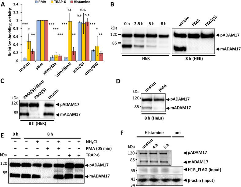
Figure 1. Effect of different stimulators on the activation and degradation of mature ADAM17.
(A) HEK293 cells were transfected with AP_IL-1RII or transfected with AP_IL-1RII and H1R. Shedding activity was measured after a 30-minute treatment with solvent/DMSO (unstim), with a stimulator (stim) or with a stimulator and an inhibitor. 100 nM PMA, 10 μM Histamine or 30 μM thrombin receptor agonistic peptide (TRAP-6) SFLLRN were used as stimulators. For inhibition cells were treated with following compounds: 10 μM metalloprotease inhibitor marimastat (Ma), 5 μM PKC inhibitor BimII, 1 μM ADAM10 inhibitor GI or 1 μM ADAM10/17 inhibitor GW. All values were normalised to the stimulated values. n = 3; *p < 0.05, **p < 0.01, ***p < 0.001. (B–F) Glycosylated proteins were enriched by precipitation with ConA-Sepharose and immunoblotted (n = 3). (B) Left panel: Influence of PMA on mature ADAM17 (mADAM17) and its proform (pADAM17) were analysed in HEK293 2.5, 5 or 8 hours after PMA stimulation. Right panel: Cells were incubated with 100 nM PMA either for the whole 8 hours (PMA) or only for 5 minutes with a washing step and incubation in media without stimulator afterwards (PMA (5)). (C) Cells were incubated with 100 nM PMA for 5 minutes and incubated for 8 hours without PMA or equally stimulated and simultaneously treated with BimII. (D) Influence of 100 nM PMA on HeLa cells. (E) Influence of PAR1 activation (30 μM TRAP-6) on ADAM17 degradation. To inhibit lysosomal degradation 50 mM NH4Cl were used. (F) HEK293 cells were transfected with H1R and were not stimulated or stimulated with histamine for 4 or 8 hours. unt: untransfected control lysate.

