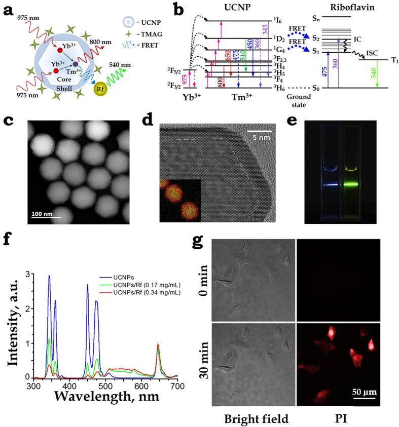
Figure 2.
(a) A schematic diagram of the core/shell UCNP, explaining the 975-nm excitation (solid red wavy lines) of Yb3+ ions in the core, which then non-radiatively transfers (dashed arrows) the energy to an Tm3+ ion that passes the energy (dashed blue wavy line) to an Rf molecule via a resonant energy transfer (RET) process. The UCNP surface is coordinated by tetramethylammonium hydroxide (TMAH). (b) Energy level diagram of a UCNP – Rf pair. Excitation at 975 nm drives Yb3+ to the excited state 2F5/2 from which it can non-radiatively transfer the energy to Tm3+ (via 3H6 → 3H5 transition, followed by the relaxation to the metastable level 3F4). Two metastable excited state 2F5/2 Yb3+ and 3F4 Tm3+ ions coalesce to drive Tm3+ to the 3F2,3 state, while an Yb3+ decays to the ground state 2F3/2. Likewise, collective energy process of 2F5/2 Yb3+ and 3F4. 3H4. 1G4 … Tm3+ drives Tm3+ to the 3H4.1G4. 1D2 …, respectively (magenta arrows). There is a probability to populate S1, S2 levels of Rf via RET or Föster RET processes from the 1G4, 1D2 levels of Tm3+. (c) High-angle annular dark-field scanning and (d) high-resolution TEM images of as-synthesised core/shell NaYF4:Yb:Tm/NaYF4 UCNPs mean-sized 75 ± 5 nm, featuring the β-crystal phase. Overlay Y (green) and Yb (red) elemental EDX mapping of UCNPs is given as a bottom left insert in (d). (e) In vitro demonstration of RET of a UCNP–Rf donor-acceptor pair. Two cuvettes filled with plain UCNPs and UCNP-FMN 0.34 mg/mL aqueous colloids illuminated with a 975-nm laser beam. The respective blue and yellow traces of photoluminescence illustrate a strong RET effect. (f) Spectra of UCNP- FMN in water under 975-nm excitation acquired at 0, 0.17 mg/mL and 0.34 mg/mL concentrations of Rf (blue, green, red curves), respectively, with the concentration of UCNPs 0.5 mg/mL. A broadband fluorescence signal from 500 nm to 620 nm ascribed to the FMN emission was a strong manifestation of the RET between UCNP and FMN. (g) Demonstration of the phototoxicity effect of UCNP-Rf pair on SK-BR-3 cells irradiated with a 975-nm laser. Phase contrast (left) and propidium iodide (PI) fluorescence (right) images of the cells before (top images) as compared to the cells after (bottom image) irradiation highlights disintegration of the cells membranes.

