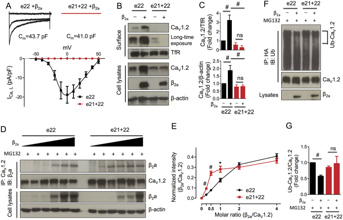
Figure 3. Characterization of exons 21 + 22-containing CaV1.2e21+22 channels.
(A) CaV1.2e21+22 channels were co-expressed with β2a and α2δ subunits in HEK293 cells. Whole cell patch-clamp recordings were performed on the cells expressing wild-type (n = 7) or CaV1.2e21+22 (n = 8) channels. (B,C) Detection and quantification of surface and total HA-CaV1.2e22 or CaV1.2e21+22 channels in the presence or absence of β2a subunit in transfected HEK293 cells (n = 4). Surface channels were biotinylated as indicated in the Methods. (D,E) Detection and quantification of β2a subunits bound to HA-CaV1.2e22 or CaV1.2e21+22 channels. β2a subunits were co-transfected with channels at different molar ratios of 0, 1/4, 1/2, 1/1, 2/1 or 4/1 (β2a/CaV1.2) in HEK293 cells treated with MG132 (2.5 μM, n = 4). (F,G) Detection and quantification of the ubiquitination levels of HA-CaV1.2e22 and CaV1.2-e21+22 channels in transfected HEK293 cells treated with MG132 (n = 3). e22, HA-CaV1.2e22 channels. e21 + 22, CaV1.2e21+22 channels. Data were shown as mean ± SEM, ns, non-significant, *p < 0.05, #p < 0.01.

