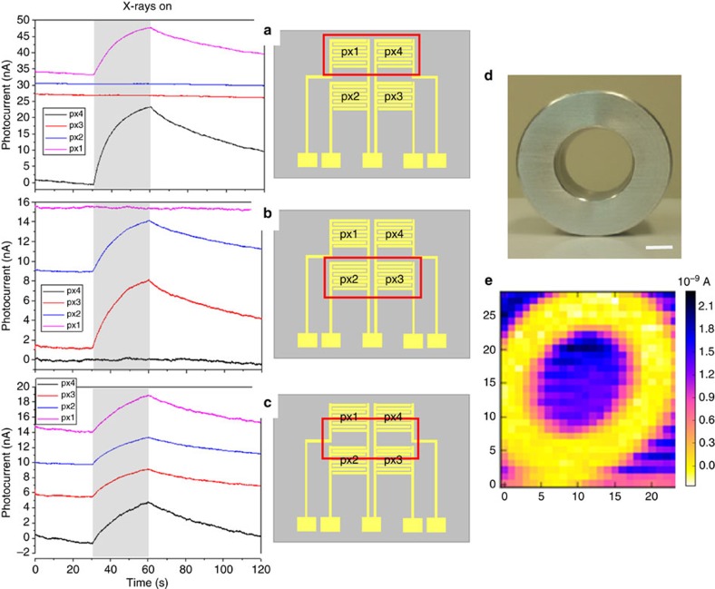
Figure 5. 2 × 2 pixel matrix organic detector.
Left: X-ray-induced current signals versus time, recorded by selectively irradiating the pixels of a 2 × 2 detector matrix. The radiation source employed is a monochromatic synchrotron X-ray beam at 17 keV with a dose rate of 28.5 mGy s−1. The pixels (with W=48 mm and L=30 μm) were all biased at 0.2 V. Note that the baseline of the four photocurrent signals are shifted in y axis for clarity. Centre: a sketch of the device is reported, with the red box indicating the region of the matrix under irradiation. In particular: in a only pixels 1 and 4 are irradiated, in b only pixels 2 and 3 are irradiated, in c all the pixels are irradiated. Right: photograph (d) and corresponding X-ray image by a single pixel device (e) of an aluminium annular ring; the scale bar is 5 mm.

