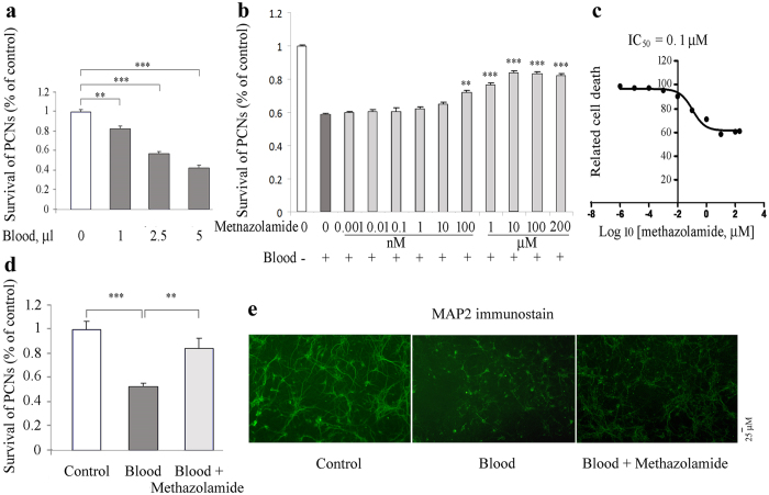
Figure 6. Methazolamide inhibits blood-induced PCNs cell death.
Blood samples are collected using retroorbital vein method in mice. Cell death of PCNs is induced by 48-hour exposure to blood: 0 (100 μl culture medium), 1 (99 μl medium), 2.5 (97.5 μl medium) or 5 μl blood (95 μl medium) (a) or 2.5 μl blood are added to 97.5 ml medium with or without a series of concentrations of methazolamide (b,c) or 10 μM methazolamide (d,e). PCNs are then fixed with 4% paraformadehyde and immunostained with MAP2 antibody. Cell death was evaluated by the MAP2 immunostaining and counting (a–d). The number of survived PCNs per well are summed from the fields counted and expressed as a percent of those present in control wells not exposed to blood. Data from 3 independent experiments are graphed, and statistically significant differences are indicated with **P < 0.01, ***P < 0.001. The extent of cell death is always normalized relative to what is measured in the absence of both death stimulus and methazolamide (white bar). The dark grey bar corresponds to the extent of cell death in response to the blood without the test drug. Light gray bars correspond to the extent of cell death with blood and methazolamide. The relative cell death results are displayed graphically. The resulting curves (plotted semilogarithmically) define the IC50 of methazolamide (c). (e) PCNs are grown in 96-well plates. Representative survival counting and images of immunohistochemistry staining of MAP2 are shown (d, e). Scale bar = 25 mm.

