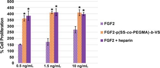
Figure 3.

Cell growth of heparin-mimicking polymer conjugates in BaF3-FR1C cells. Incubation of 20 000 cells/well in 96-well plate with FGF2 or the heparin-mimicking polymer conjugates in the absence or presence of 1 μg/mL of heparin was carried out for 48 h. CellTiter-Blue assay was performed to quantify the extent of cell growth. Data was normalized to the blank medium group, which was set at 100%. Each sample contained four replicates and the experiment was repeated three times. Error bars represent standard error of the mean (SEM). Statistical analysis was done using Student’s t test. * p < 0.01 compared to FGF2.
