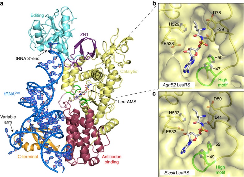
Figure 3. Structure of the AgnB2·tRNALeu·Leu-AMS complex.
(a) X-ray structure of wild-type AgnB2·tRNALeu·Leu-AMS complex with 3′-tRNALeu (blue) bound in the editing site (PDB Code—5AH5). The catalytic, editing, anticodon binding and C-terminal domains are displayed in yellow, cyan blue, red and gold, respectively. Leu-AMS bound to the synthetic active site is represented in a stick structure and the catalytic KQSKS and HIGH loops are highlighted in green. (b,c) Comparison of wt AgnB2·tRNA·Leu-AMS complex and LeuRSEc· tRNALeu·Leu-AMS complex binding site residues. The surface of the catalytic active site of the protein is depicted in yellow with Leu-AMS bound (stick structure).

