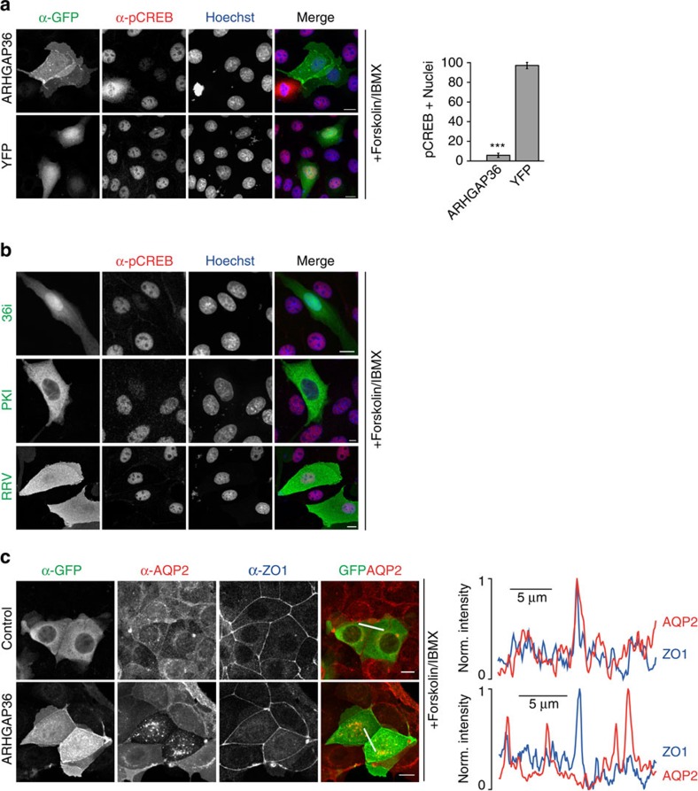
Figure 7. ARHGAP36 suppresses PKA signalling.
(a) MDCK cells expressing YFP-ARHGAP36 or YFP control in low-serum conditions were treated with 10 μM Forskolin and 100 μM IBMX for 20 min, fixed and subjected to immunofluorescence using antibodies against GFP and phospho-CREB. Images were collected by confocal microscopy. Scale bars, 10 μm. Quantitative analysis of nuclear phospho-CREB staining in cells expressing the indicated constructs (n>100 for each of three independent experiments, shown as mean±s.e.m., ***P<0.001, Student's T-test). (b) As in a, except cells were transfected with Cherry-36i, CFP-PKI or CFP-ARHGAP36-RRV. (c) MCD4 cells stably expressing aquaporin-2 (AQP2) were transfected with YFP-ARHGAP36 or YFP control in low-serum conditions. Twelve hours post transfection, cells were treated with 10 μM Forskolin for 20 min. Fixed cells were subjected to immunofluorescence using antibodies against GFP, AQP2 and the tight junction protein ZO-1, to visualize the plasma membrane at cell–cell contact sites. Images were collected by confocal microscopy. Scale bars, 10 μm. Line scan fluorescence intensity profiles are shown on the right. In red: AQP2, in blue: ZO-1.

