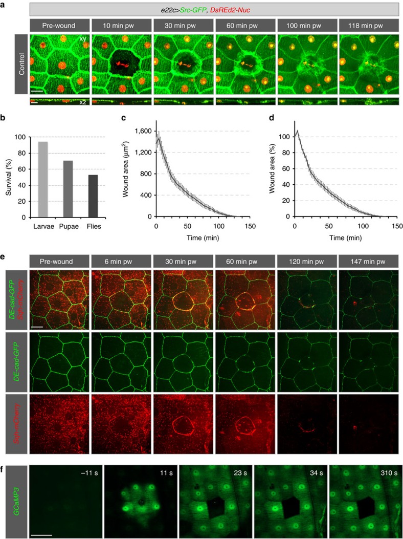
Figure 1. Healing of single-cell epidermal wounds in 3rd instar larvae.
(a–d) Heterozygous e22c>Src-GFP,DsRed2-Nuc (e22c-Gal4, UAS-Src-GFP, UAS-DsRed2-Nuc/+), (e) recombinant DE-Cad-GFP, Sqh-mCherry and (f) e22c>GCaMP3 (e22c-Gal4, UAS-GCaMP3) early third instar larvae were immobilized and wounded by laser. (a) Projections (xy) and cross-sections (xz; stacks) of the wounded epidermis from a time-lapse series of a single-cell wound that was closed by 118 min. (b) Survival rate of larvae after wounding. After single-cell wounding 94% of the larvae survived 48 h, 71% pupated and 59% eclosed as adult flies. (c–d) Quantitative analysis of wound healing (n=10 larvae). Data given as mean±s.e.m. (c) Changes in absolute wound area (μm2) and (d) in relative wound area (%). After single-cell ablation, the wound area first expanded for 6±1 min. 50±4% of the wound area was covered within ∼23 min and the remaining 50% closed within ∼95 min. (e) Actomyosin dynamics of single-cell wound healing of n=6 larvae. At 6±1 min an actomyosin cable started to form and was completed at 10–14 min and maintained throughout wound healing. (f) Response of the Calcium sensor GCaMP3 (green) to wounding. GCaMP3 is ubiquitously expressed in the epidermis. After wounding a calcium wave spreads from the wounded area. Scale bars, (a,e) 20 μm, (a) in xz section: 10 μm, (f) 30 μm. pw: post-wounding, DE-Cad: Drosophila E-Cadherin, Sqh: spaghetti squash, the Drosophila non-muscle myosin II regulatory light chain.

