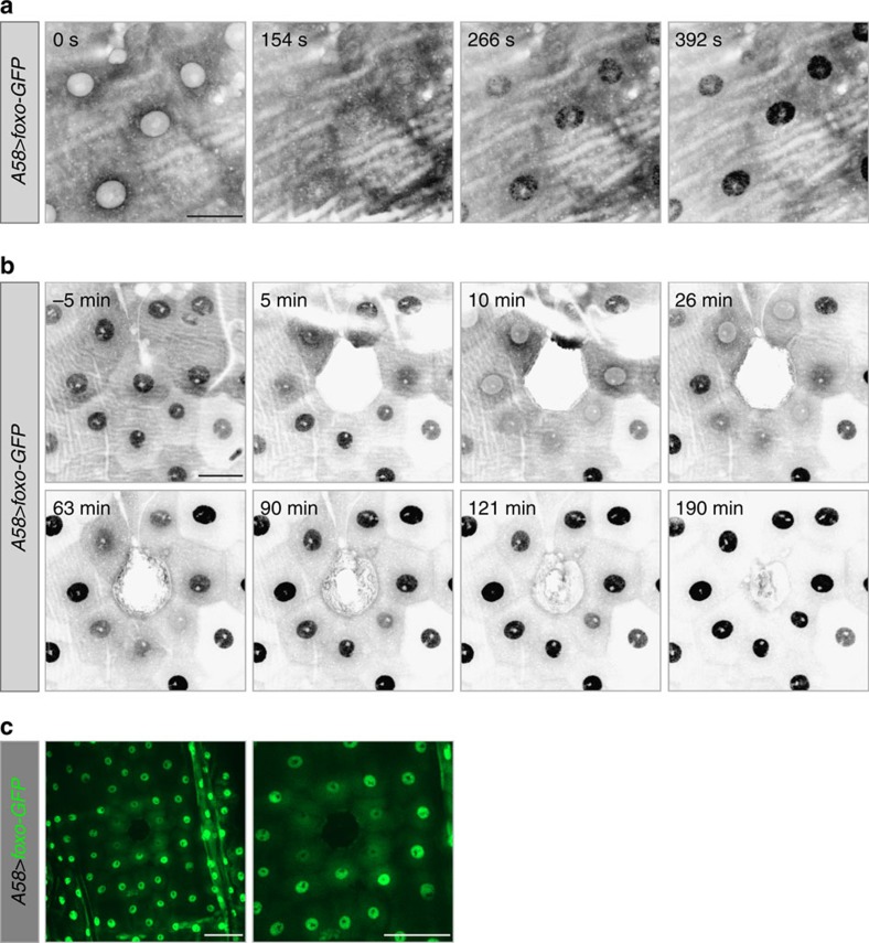
Figure 6. Subcellular distribution of overexpressed FOXO in the epidermal cells.
(a–c) FOXO distribution before and after wound healing in larvae overexpressing foxo-GFP in the epidermis (A58>foxo-GFP). (a) When larvae are first mounted for spinning disk confocal microscopy (488 nm laser), FOXO-GFP (grey) is present at higher levels in the cytoplasm than in the nucleus. FOXO-GFP then begins to be enriched in the nucleus, reaching maximal nuclear levels after ∼350–400 s; this also occurred when the larvae were observed only with epifluorescence, though at a slower rate. (b) On wounding most of the nuclear FOXO (grey) was shuttled into the cytoplasm (10–26 min). During the course of wound healing FOXO then again accumulated in nuclei, with high levels reached by ∼60–90 min. (c) Lower magnification view of subcellular distribution of FOXO-GFP (green) 12 min after wounding. Cells at a distance from the wound edge did not respond to wounding and FOXO-GFP remained nuclear. Scale bars, (a) 20 μm, (b) 30 μm and (c both panels) 60 μm.

