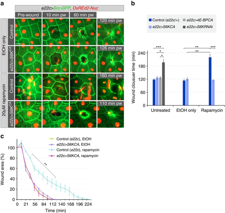
Figure 7. S6K activation suppresses rapamycin-induced wound healing delay.
(a–c) Wound healing in larvae expressing constitutively active S6K (S6KCA). All larvae expressed Src-GFP (green) and DsRed2-Nuc (red). (a) Time-lapse images of wound healing in L3 larvae. (b) Average time of wound closure for genotypes shown in a and Supplementary Fig. 12A: Untreated larvae: 118±5 min for control, 126±7 min for e22c>S6KCA, 127±5 min for e22c>4E-BPCA and 196±12 min for e22c>S6KRNAi; larvae treated only with EtOH 114±6 min for control and 120±5 min for e22c>S6KCA; larvae treated with 20 mM rapamycin in EtOH: 220±12 min for control and 116±2 min for e22c>S6KCA. (c) Time course of changes in relative (%) wound area. (b,c) Mean±s.e.m., (b) one-way ANOVA with post hoc test and (c) two-way ANOVA with post hoc test (comparing control and Rapamycin treatment). *P<0.01, **P<0.001 and ***P<0.0001, n=3–5 larvae each genotype and each treatment. Scale bar, (a) 20 μm. Transgene genotypes of larvae: Control (e22c-Gal4/+), e22c>S6KCA (e22c-Gal4; UAS-S6KSTDETE), e22c>4E-BPCA (e22c-Gal4; UAS-4E-BPCA) and e22c>S6KRNAi (e22c-Gal4; UAS-S6KRNAi).

