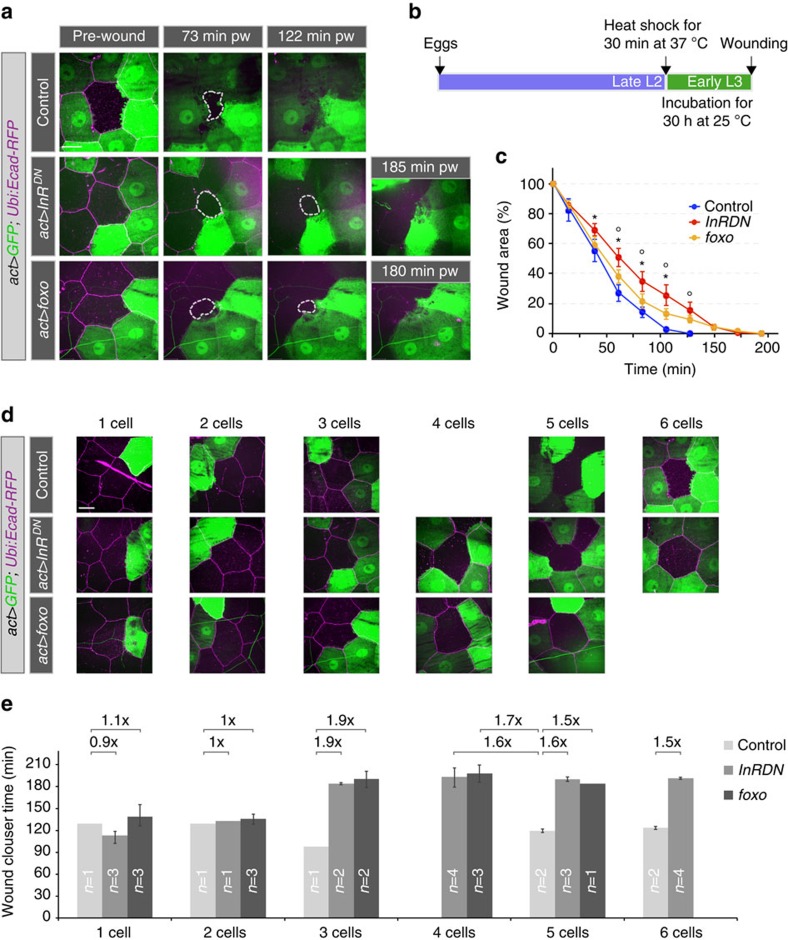
Figure 8. Clonal analysis of the requirement for insulin signalling.
(a–e) Wound healing in larvae with epidermal clones of modified InR and FOXO activity. Ubiquitously expressed DE-Cad-RFP was used to visualize all cells in the epidermis. (a) Time-lapse images of wound healing in L3 larvae. (b) Schematic for timing of transgene expression. (c) Kinetics of wound closure in larvae expressing InRDN or FOXO in 3–6 cells surrounding a wounded cell. Time course of changes in relative wound area. Mean±s.e.m. one-way ANOVA with post hoc test *P<0.01 (control versus InRDN) and °P<0.01 (control versus foxo), n=3–5 larvae for each genotype. (d) Examples of single-cell wounds with varying numbers of cells expressing InRDN or FOXO or only GFP as their neighbours. (e) Average time of wound closure for mosaic genotypes shown in d. Mean±s.e.m., ‘n' is the number of larvae analysed in each case. Scale bars, (a,d) 20 μm. Transgene genotypes of larvae: Control (hsflp; act>y+>Gal4, UAS-GFP/+; DE-Cad-RFP/+), act>InRDN (hsflp; act>y+>Gal4, UAS-GFP/UAS-InRDN; DE-Cad-RFP/+), act>foxo (hsflp; act>y+>Gal4, UAS-GFP/UAS-3xflag-foxo; DE-Cad-RFP/+).

