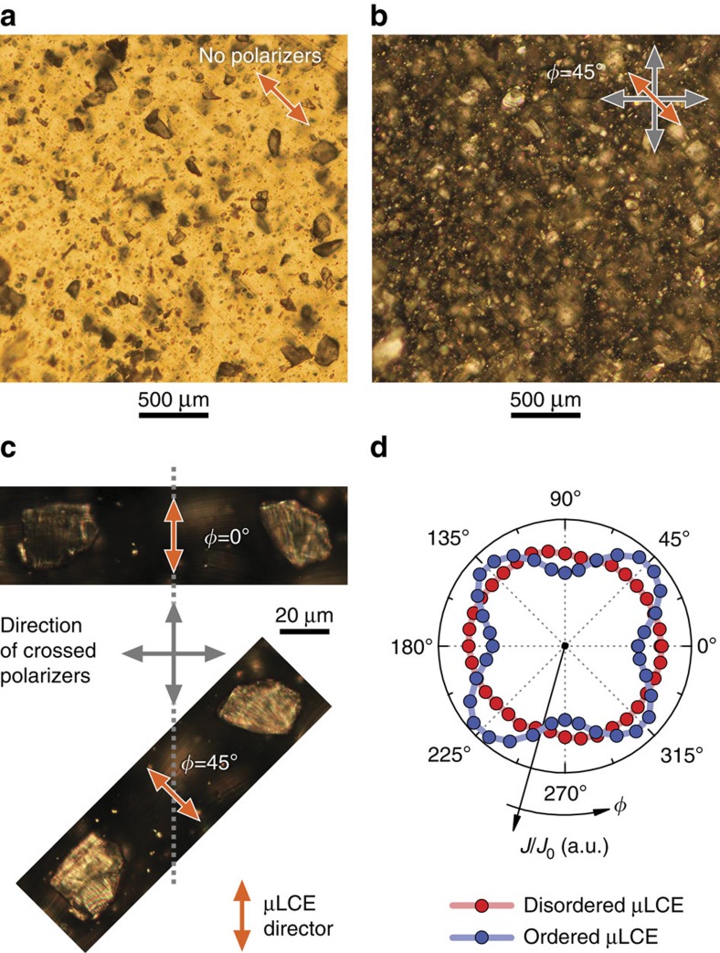
Figure 3. Structure of PDLCE as seen under polarizing optical microscope.
(a) Without polarizers, the isotropic matrix transmits light, whereas LCE microparticles are seen as dark spots. (b) Under crossed polarizers, the light passing through the matrix is blocked, whereas LCE microparticles do transmit light due to their anisotropic nature. (c) Two microparticles compared at different orientations with respect to polarizers. The particles are dark when their directors are aligned with one of the polarizers (ϕ=0°) and bright at ϕ=45°. PDLCE-A sample, cut into 0.1 mm thick slice, with low, 5% wt concentration of μLCE-A, was used for microscopy investigations. (d) Polar plot of the sample's transmittance J/J0 demonstrating the optical anisotropy of the aligned (ordered) sample, arising from the alignment of μLCE particles.

