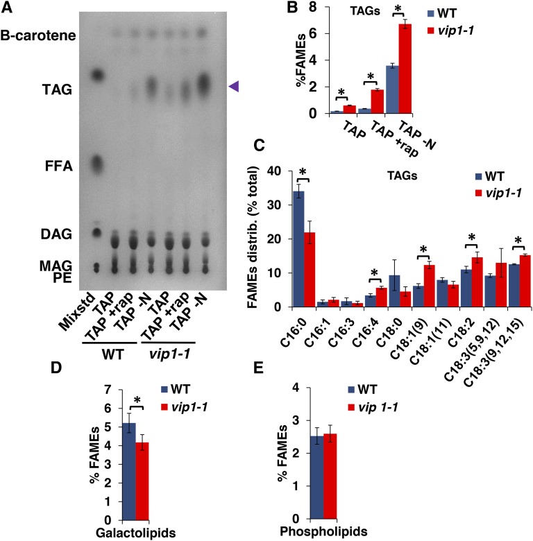
Figure 6.
Increased TAG and Decreased Galactolipids in vip1-1.
(A) TLC separation and iodine staining of different lipid classes from total lipids of wild-type and vip1-1 cells growing in TAP, TAP supplemented with rapamycin for 12 h (+rap), or transferred to TAP-N for 12 h. The left-most lane contains a mixture of standards (Mixstd) whose constituents are abbreviated as follows: PE, phosphatidyl ethanolamine; MAG, monoacylglycerol; DAG, diacylglycerol; FFA, free fatty acids; TAG, triacylglycerol. The identification of β-carotene in the fastest migrating position was done separately using a purified standard. Blue arrowhead on right indicates migration of TAGs.
(B) Quantitation of FAMES derived from purified TAGs and normalized by dry cell weight for strains shown in (A). Errors bars indicate sd from five replicates.
(C) Relative distribution (percent total) of acyl group types from TAG fraction of wild-type and vip1-1 strains.
(C) and (D) Percentage of FAMEs in galactolipid and phospholipid fractions purified from the wild type and vip1-1 and normalized by dry cell weight. Error bars indicate sd from at least three biological replicates. Asterisks represent significant differences (P < 0.05) between means evaluated using Student’s t test.

