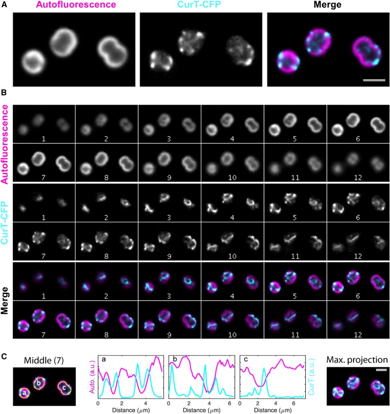
Figure 8.
Live-Cell Imaging of the curT-CFP Strain by Fluorescence Microscopy.
In the curT-CFP strain, the native curT gene has been replaced by a CFP-tagged version resulting in the expression of the CurT-CFP fusion protein under the control of the native promoter.
(A) Close-up views of the mid-cell plane in the CFP and far-red autofluorescence channels shown in slice 7 in (B).
(B) Synechocystis 6803 cells expressing CurT C-terminally fused to the mTurquoise2 variant of CFP from its native chromosomal promoter. The two imaging channels are displayed as z axis montages (auto-scaled contrast, step size 250 nm) in grayscale and color composite configuration. The thylakoid autofluorescent channel as imaged in far-red fluorescence is depicted in magenta and CurT signal as imaged in cyan fluorescence is depicted in cyan.
(C) Fluorescence intensity profiles of the two channels along the autofluorescent peripheral cell “ring” are shown for slice 7. The maximum projection of the entire Z montage is also shown. Bars (light gray) = 2 µm.

