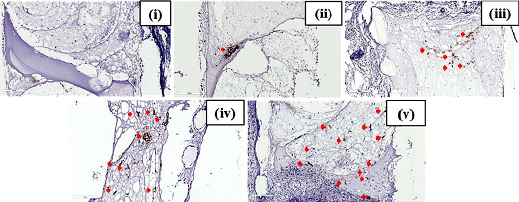
Figure 3.
Human specific CD-31 staining of in vivo implanted Poly(propylene fumarate)/Fibrin Scaffolds. The control group (C) did not show any positive staining for endotheial cells depicted by brown staining (i). The no-preculture (ii), 1 week preculture (iii), 2 week pre-culture (iv) and 3 week preculture (v) groups show an increase in vascular area from one group to the next as depicted by brown stained human endothelial cells (indicated by red arrows) mostly forming vessel-like structures in the 1P, 2P and 3P groups as compared to that of NP group, showing positive stained cells accumulated in a single area indicating the presence of spheroids. Scale bar of each image is 300µm.

