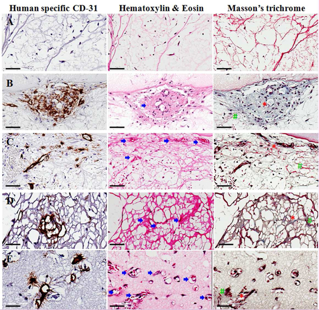
Figure 4.
Image showing the presence of vascular areas via staining of serial sections for human specific CD-31 staining, Hematoxylin & Eosin (H&E) as well as Masson's trichome staining in control (A), no pre-culture (NP) (B), 1 week pre-culture (C), 2 week pre-culture (2P) (D) and 3 week pre-culture (3P) (E) groups. All the groups show large amount of cells except the control group. The CD-31 immunohistochemistry-stained perfused vascular areas (indicated by blue arrow) can be seen via H&E and Masson’s trichrome stain. Masson’s trichrome staining also reveals structures such as collagen fibrils (indicated by red ‘*’ symbol) as well as fibrin (indicated by green ‘#’ symbol). The scale bar in each image represents 70µm.

