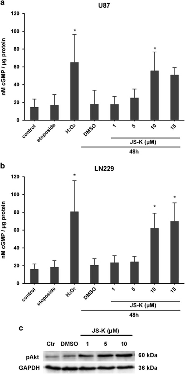
Figure 3.
Dose-dependent increase of intracellular cGMP in U87 (a) and LN229 (b) cells mediated by JS-K after 48 h as well as in necrotic controls exposed to H2O2 (3 mM). cGMP level remained stable after induction of apoptosis by etoposide (10 μM). Asterisks (*P<0.05, **P<0.01, ***P<0.001) indicate significance compared to untreated controls. Western blot analysis for pAkt in LN229 (c) cells show increase of phosphorylation after exposure to JS-K for 48 h compared to control. The blot is representative for three independent experiments

