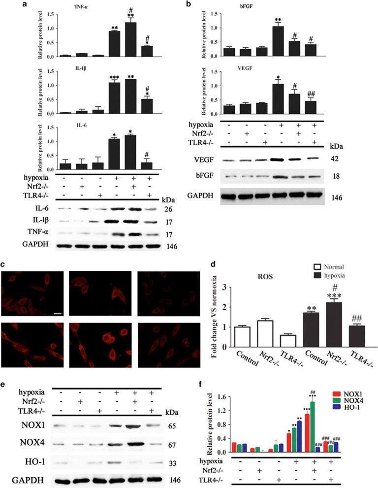
Figure 5.
Effect of Nrf2 and TLR4 on the expression of inflammatory cytokines, growth factors, and intracellular and mitochondrial ROS generation in ADSCs. ADSCs were isolated from WT, Nrf2−/− or TLR4−/− mice and exposed or not to hypoxic conditions. (a and b) TNF-α, IL-1β, IL-6, VEGF and bFGF expression in ADSCs was measured by western blotting (n=5). (c) Micrographs of ADSCs labeled with dihydroethidium to detect ROS (magnification, × 40). Scale bar: 20 μm. (d) Quantification of mitochondrial DCF fluorescence intensity in ADSCs (n=5). (e) NADPH oxidase (NOX) expression was measured by western blotting (n=5). (f) Relative protein expression and fold change of NOX1, NOX4 and HO-1 relative to GAPDH (n=3). *P<0.05, **P<0.01, ***P<0.001 versus the control group, and #P<0.05, ##P<0.01, ###P<0.001 versus the hypoxia control group

