Figure 2.
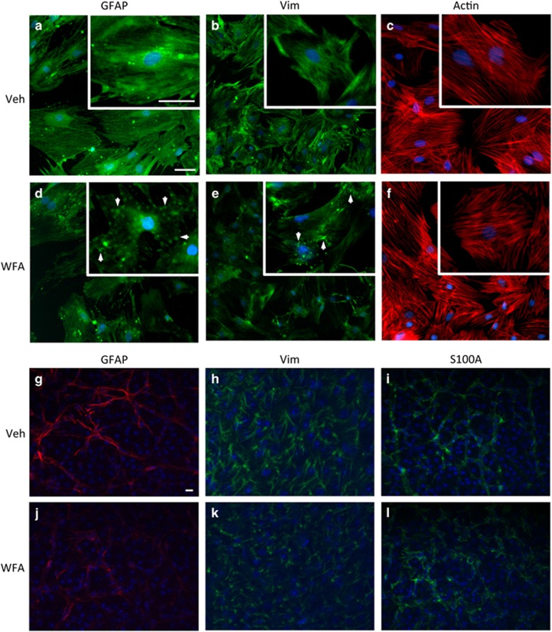
WFA treatment inhibits IF dynamics. (a–f) Treatment of cultured retinal astrocytes with 2.0 μm WFA resulted in reduced staining for GFAP (a and d), and vimentin (vim; b and e), compared with control treated cells after 8 h. At higher magnifications (insets), filamentous staining for GFAP and vimentin was disrupted, resulting in formation of IF aggregates (arrows). In comparison, filamentous actin remained unaffected (c and f). (n=3, scale bars indicate 100 μm. Identical exposures were used for all treatments). (g–l) WFA treatment in vivo similarly disrupts GFAP and vimentin in retinal flatmounts, but not S100A. (n=3, scale bars indicate 100 μm. Identical exposures were used for all treatments)

