Figure 3.
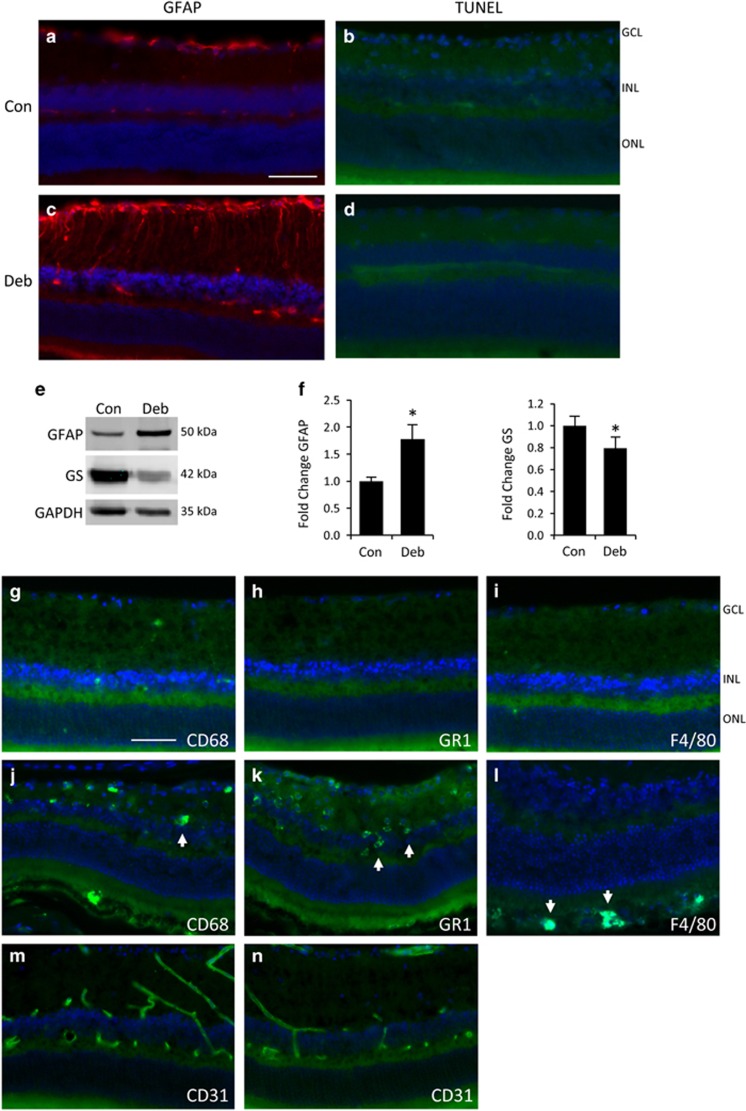
Rapid retinal glial reactivity is induced by corneal injury in the absence of cell death or inflammation. (a and b) Non-debrided control eyes show no evidence of gliosis or cell death. (c and d) Mechanical debridement of the corneal epithelium in the contralateral eye results in strongly increased GFAP staining in retinal astrocytes and Müller glia by 7 days (c), but no evidence of cell death or disrupted morphology (d). (e) Western blots from whole retina lysates show increased GFAP and decreased GS. (f) Densitometry and statistical analyses from multiple blots confirming the changes in e (bars represent SE, *P<0.05, n=3 animals). (g–i) Staining of debrided retinas for activated microglia, neutrophils and macrophages, was largely negative with CD68, GR-1 and F4/80 antibodies, respectively. (j–l) Corresponding antibody-positive controls from eyes treated with 150 mM NaOH. (m and n) There was no change in retinal blood vessel staining with the endothelial marker CD31 between control (m) or debrided eyes (n). (Scale bar indicates 50 μm, Con; control, Deb; debrided.)

