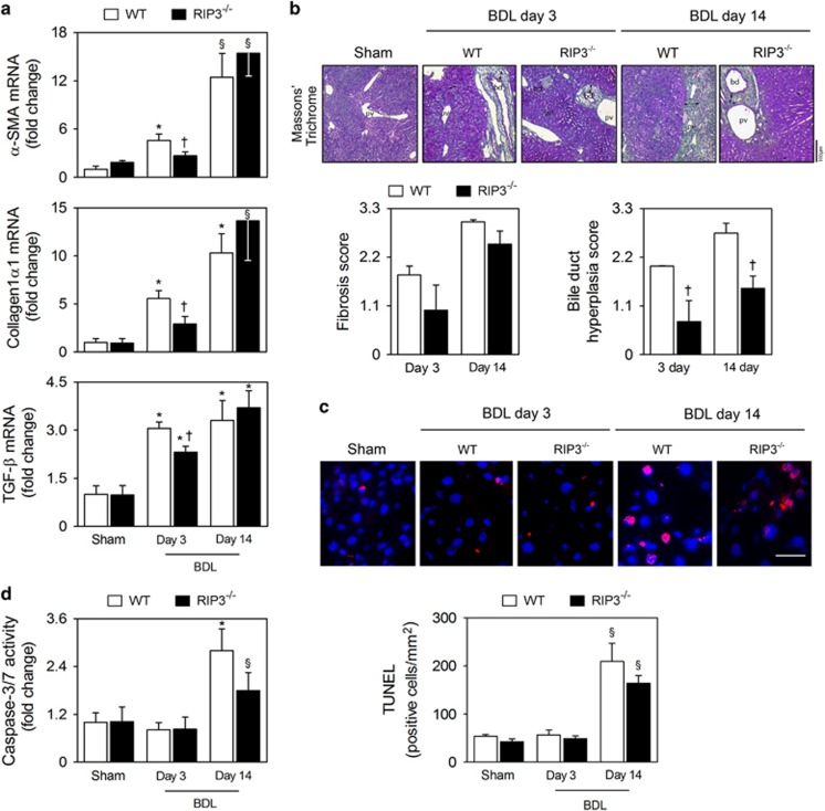
Figure 4.
Deletion of RIP3 does not improve BDL-induced fibrosis and apoptosis. C57BL/6N WT and RIP3−/− mice were subjected to sham or BDL surgical procedures and killed at days 3 and 14. (a) qRT-PCR analysis of α-SMA, collagen-1α1 and TGFβ in mouse liver. Results are expressed as mean±S.E.M. fold change of 7–10 individual mice. (b) Representative images of Masson's Trichrome-stained liver sections (top). Periportal fibrosis and bile duct hyperplasia were scored as described in Materials and Methods section (bottom). Results are expressed as mean±s.e.m. fold change of 4–5 individual mice. (c) TUNEL staining of liver tissue sections. Nuclei were counterstained with Hoechst 33258 (blue). Scale bar, 30 μm (left). Histograms show the quantification of TUNEL-positive cells/mm2 (right). Results are expressed as mean±S.E.M. fold change of 4–5 individual mice. (d) Caspase-3/7 activity assay. §P<0.05 and *P<0.01 from sham-operated mice; †P<0.05 from BDL WT mice at respective time-point

