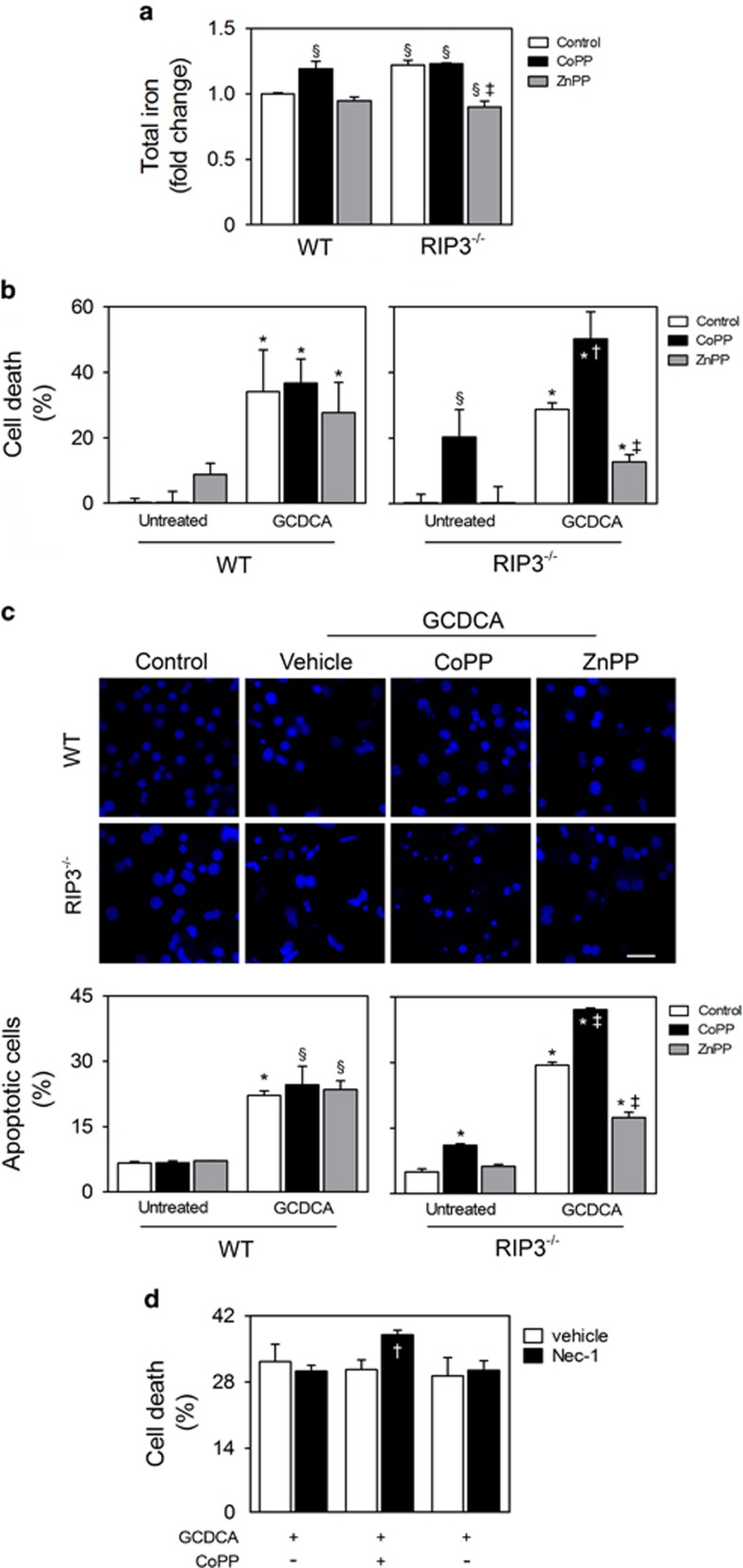
Figure 6.
HO-1 is involved in iron accumulation and cytotoxic effects of GCDCA in RIP3-deficient primary mouse hepatocytes. Primary mouse hepatocytes isolated from WT and RIP3−/− C57BL/6N mice were treated with CoPP (10 μM), ZnPP (15 μM) or vehicle control. After 1 h, cells were exposed to either GCDCA (50 μM) or vehicle control. After 24 h, cells were harvested for total iron or immunoblotting. Cell death assays were performed after 48 h. (a) Total iron levels in whole cells were measured as described in Materials and Methods section. Values were normalized with total protein concentration. (b) Percentage of general cell death in primary mouse hepatocytes as assessed by LDH activity assay. (c) Apoptotic cells detected by Hoechst staining. Results are expressed as percentage of apoptotic cells (bottom). Representative images of untreated WT and RIP3−/− primary hepatocytes and incubated with GCDCA, GCDCA plus CoPP or GCDCA plus ZnPP are shown. Scale bar, 30 μM. (d) Primary mouse hepatocytes isolated from WT and RIP3−/− C57BL/6N mice were treated with Nec-1 (100 μM) or vehicle control. After 1 h, cells were exposed to GCDCA (50 μM) for 48 h. Percentage of general cell death was as assessed by the LDH activity assay. Results are expressed as mean±S.E.M. fold change or percentage from three independent cultures from each genotype. §P<0.05 and *P<0.01 from Control; †P<0.05 and ‡P<0.01 from respective control

